By: Jason H. Goodchild, DMD
Whether the clinical situation is being captured conventionally by impression material or via digital scan, soft tissue management during crown and bridge procedures is vitally important to making an acceptable impression. A study from Samet and colleagues found that 89% of impressions made had one or more observable errors; common errors included voids, bubbles, and flow problems. (J Prosthet Dent 2005;94:112-7.) Mitigating some of these errors definitely includes improved tissue management, and in particular hemostasis and drying of the area before impression material is placed or digital capture occurs.
Prior to capturing a final impression, many practitioners have historically used mechanical retraction through the placement of cord, alone or in combination with a chemical agent to promote hemostasis and retract tissue (e.g., aluminum chloride, ferric sulfate, racemic epinephrine). In one early study of North American dentists, it was found that 95% of respondents routinely used gingival retraction cord during crown and bridge impressions, (J Prosthet Dent 1985;53(4):525-31.) and in another study 98% of prosthodontists reporting using cord. (J Prosthodont 1999;8:163-70) To allow enough material to create an undistorted impression, a 0.2mm space must be created between tooth and gingiva. This minimum space requirement is intended to allow the impression material to flow into the sulcus and not exceed its tear-strength capability, preventing the chance for remnants to be left behind that could cause gingival irritation and inflammation. This minimum space requirement for impression material is diminished in part for digital impressioning, however interfering tissue must still be displaced or removed in order to properly record the entirety of the preparation.
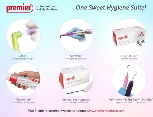
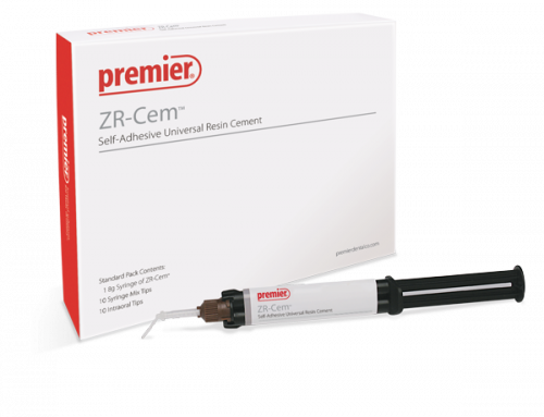
It is well known that placing cord creates an acute tissue injury that in some cases can lead to post-operative discomfort, inflammation, and marginal recession. (J Prosthodont 2006;15(2):108-12.) Recently, product manufacturers have introduced retraction paste as a less traumatic means of achieving hemostasis and local retraction (e.g., Traxodent© [Premier Dental Products Company]). Even though retraction paste has been shown to create temporary gingival inflammation, it is considered less traumatic when compared to placing retraction cord and is as effective in promoting hemostasis. (Res J Biol Sci 2009;4(3):335-9. & JADA 2003;134(11):1485.)
Generally clay-based to absorb moisture and coupled with an astringent, retraction pastes are designed to be placed into and around the gingival sulcus and within several minutes produce hemostasis and drying. When used with a compression cap (a cylindrical, dense cotton pellet) and direct pressure, retraction paste can also provide excellent tissue retraction. Traxodent® is a Hemodent® Paste Retraction System that features a functionalized proprietary clay. Compared to other kaolin-based clay systems, Traxodent’s clay delivers improved ion exchange (of the astringent) and because of its surface area provides “swelling” capacity for fluid control and drying.
Traxodent contains 15% aluminum chloride and comes in either pre-packaged syringes utilizing bendable metal tips, or unit doses with a slender plastic tip that fit into an autoclavable dispenser. Information from the Manufacturer indicates that “Traxodent can be used in virtually any clinical situation in which control of bleeding is required.”(Traxodent Directions for Use) There is essentially no learning curve to using Traxodent, the material is simply dispensed into the area around the prepared tooth (with or without retraction cord) followed by having the patient bite on a Retraction Cap (Premier). After two minutes, the paste is removed by thoroughly rinsing the area with water. After gentle drying, the tissue should appear free of moisture and blood and ideally prepared for the final crown and bridge impression.
Finally, because getting the impression right the first time is critical to office efficiency there are some practitioners that “throw everything at it.” Meaning, gingival retraction cord with paste may be used to add extra insurance that the first attempt will be successful. (Clinicians Report 2015;8(3):1-9) In these situations there is no downside except a small increase in material costs and upfront time, but if it means better and more successful impressioning – go for it!
Case example highlighting the techniques

Occlusal view of tooth No. 30. The tooth has had root canal therapy, and a post & core build-up

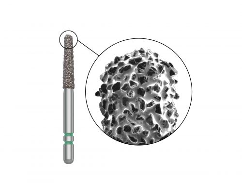
Preparation of tooth No. 30 for a PFM is completed using Two Striper® diamond burs.

Occlusal view of the completed preparation for tooth No. 30.

To control moisture and create hemostasis around the finished preparation, Traxodent® retraction paste is dispensed around the entire tooth.

The patient was instructed to bite down on a Retraction Cap for 2 minutes, followed by water rinsing to remove the paste

Occlusal view of tooth No. 30 after thorough rinsing, note the excellent hemostasis and dry field.

Completed final impression of tooth No. 30 using Impregum™ (3M™ ESPE™) and T-LOC™ Triple Tray®

Occlusal view of final PFM crown No. 30

Buccal view of final PFM crown No. 30.
®Two Striper is a registered trademark of the product manufacturer, Abrasive Technology, Inc. / ™Impregum, 3M and ESPE are not trademarks of Premier Dental® / Images courtesy of Shalom Mehler, DMD / Laboratory work by Ceramtek Dental Studio, Fair Lawn, N.J.


